About Joint use of Research Facilities
We actively promote the joint use of research equipment to enhance collaborative research efforts with both internal and external stakeholders.
For more information, please refer to the General Guidelines for the Use of Shared Research Equipment at our university (PDF).
If you wish to make use of our shared research equipment, kindly complete the Joint Use of Research Equipment Application Form (PDFform) and submit it to the contact information provided below.
| Contact | Dr. Atsuko Matsushita |
|---|---|
Research Facilities
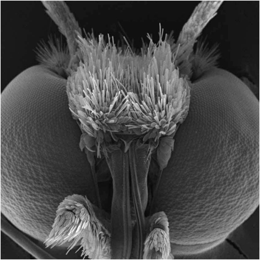
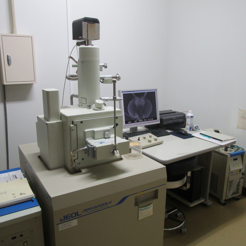
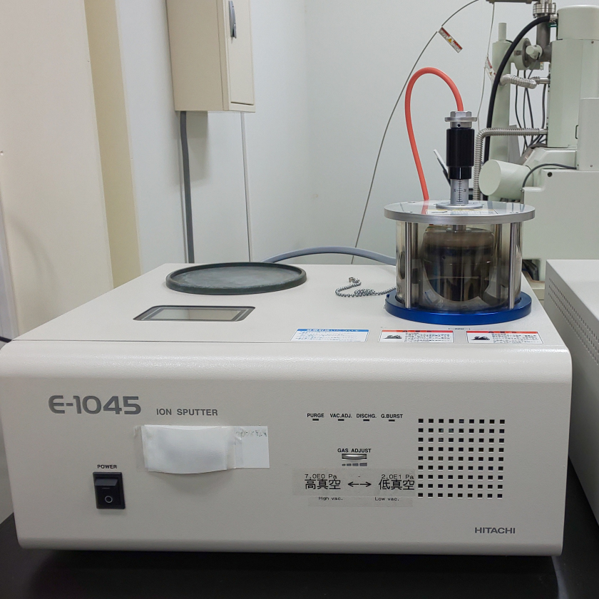
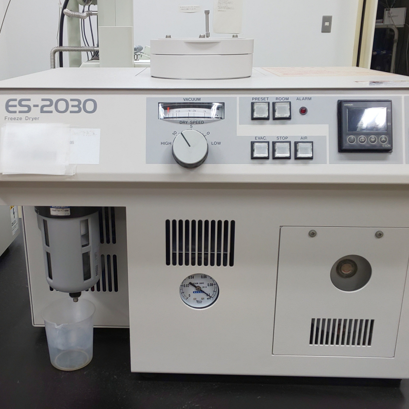
1. Scanning Electron Microscope
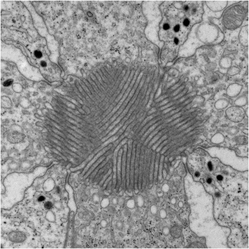
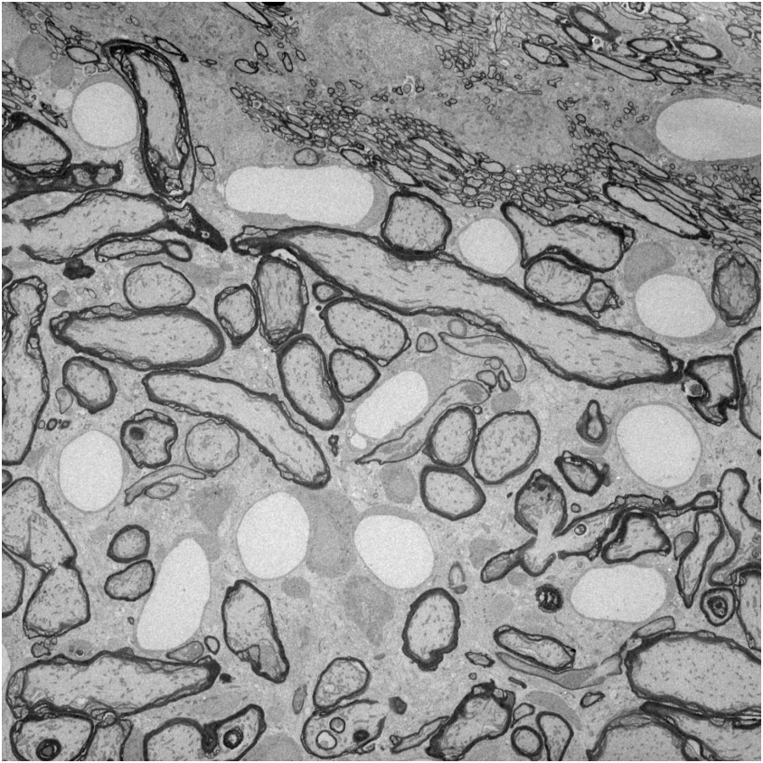
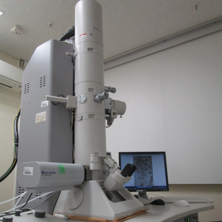
2. Transmission Electron Microscope
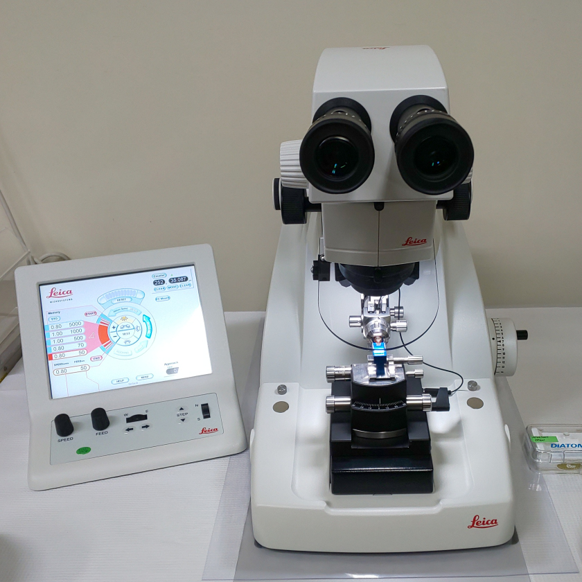
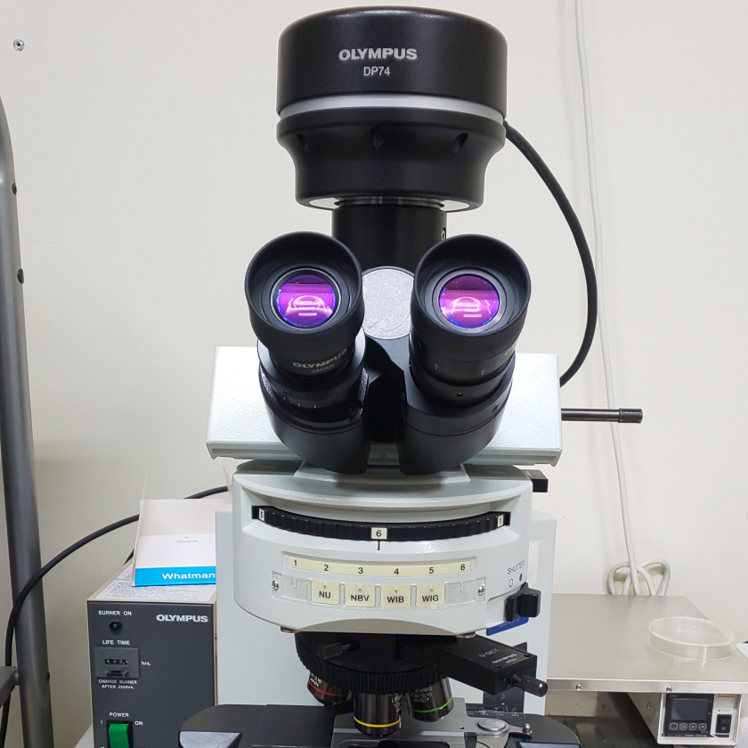
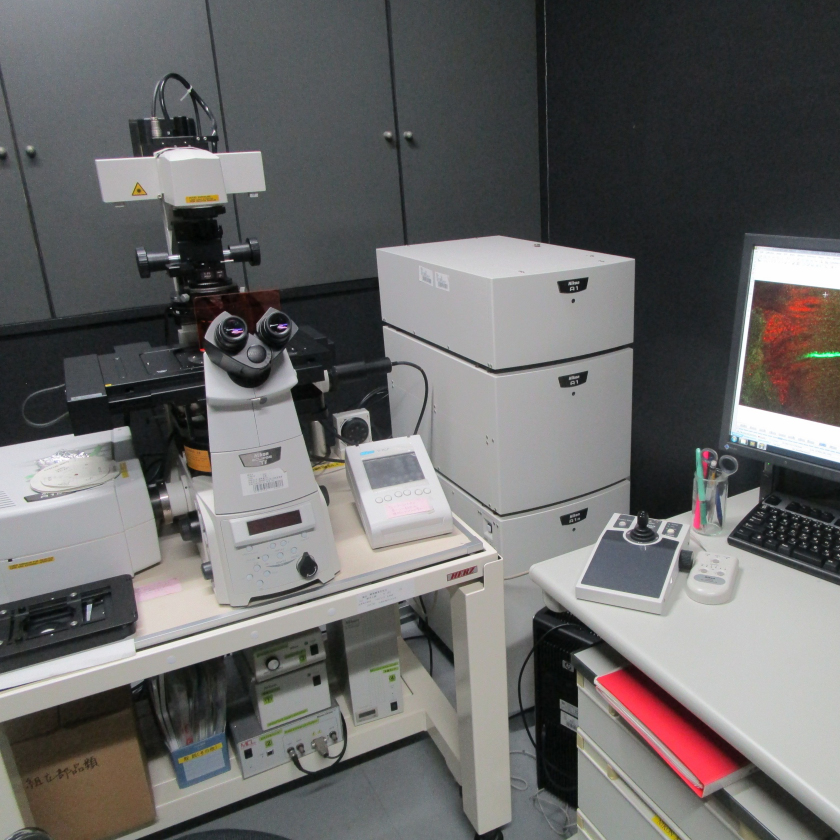
Confocal Laser Scanning Microscope

Related Page
Collaboration
→